
ECG vs. Echocardiogram: Which Heart Test Do You Actually Need?
Table of Contents
Toggle‘ECG vs. Echocardiogram: Which Heart Test Do You Actually Need?’ – an ultimate guide by Dr. Kuntal Bhattacharya, Senior Interventional Cardiologist, Narayana Health – RN Tagore Hospital, Kolkata
Every single day in my clinic in Mukundapur, Kolkata, I sit down with patients who walk in feeling anxious, confused, and overwhelmed. Often, they come to me because of a sudden episode of chest discomfort, an unexpected wave of dizziness, or breathlessness while climbing a simple flight of stairs. Inevitably, the first words out of their mouth are: “Doctor, I need my heart checked.”
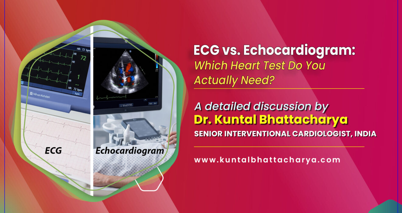
When we begin discussing the evaluation process, medical terms are quickly introduced. Two of the most foundational, everyday diagnostic tools we prescribe in modern cardiology are the ECG (Electrocardiogram) and the Echocardiogram (frequently called an “Echo”).
Because these two names sound remarkably similar, I find that many patients—and even some families—use them interchangeably. People often assume that if they have already undergone one of these tests, they have effectively checked all the boxes for their heart health.
However, nothing could be further from the truth. While both tests are entirely non-invasive, safe, and painless windows into your cardiovascular system, they look at completely different aspects of your heart.
An ECG vs. Echocardiogram comparison is never about deciding which test is “superior.” Instead, it is about understanding which diagnostic tool is specifically engineered to answer the clinical questions your heart is asking. In this comprehensive guide, I want to walk you through how these two vital tests work, what they show, and how we as cardiologists determine which one you actually need.
1. Introduction: The Language and Mechanics of Your Heart
Your heart is a remarkable, tireless muscular pump roughly the size of your clenched fist. Beating around 100,000 times every single day, it circulates about 2,000 gallons of blood through a vast network of blood vessels spanning tens of thousands of miles. To keep this intricate biological engine running smoothly without invasive measures, we rely heavily on non-invasive diagnostic tools that can listen to, record, and image the heart.
In clinical cardiology, these tests form the bedrock of effective diagnosis and treatment. Whether you are visiting a healthcare hub in South Kolkata, Salt Lake, or anywhere across West Bengal, initial cardiac evaluations almost always start right here:
- The ECG evaluates the electrical wiring of your heart.
- The Echocardiogram evaluates the mechanical structure and pumping efficiency of your heart.
My goal in writing this guide is to clear up the confusion, ease any pre-test anxiety you might feel, and help you understand your own health journey.
2. What is an ECG (Electrocardiogram)?
Defining the Test
An Electrocardiogram (commonly referred to as an ECG or EKG) is a rapid, completely painless test that records the electrical signals traveling through your heart muscle. Every single heartbeat is triggered by a tiny electrical impulse originating in your heart’s natural pacemaker (the sinus node). As this electrical wave sweeps across the heart tissue, it commands the muscle chambers to squeeze and relax in precise coordination, pumping blood forward.
An ECG machine does not send any electricity into your body. Think of it simply as a highly sensitive microphone listening to the electrical currents your heart generates naturally, printing them out as wavy lines on graph paper or displaying them on a digital screen.
How an ECG is Performed: Step-by-Step
If I order an ECG for you during your consultation, here is what the procedure looks like:
- Preparation: You will be asked to strip to the waist and lie down comfortably on an examination table. Small, sticky sensor patches called electrodes will be placed on your chest, arms, and legs. (For men with dense chest hair, a quick trim in those tiny spots may be necessary to ensure the stickers make proper contact with the skin).
- Hooking Up: Wires (leads) from the ECG machine are clipped onto these electrodes.
- The Recording: You will be asked to lie completely still, relax, and breathe normally for about 10 to 30 seconds. You won’t feel a thing.
- Completion: The technician unhooks the wires, removes the sticky patches, and prints out the tracing. The entire process takes less than 5 minutes.
What Conditions Does an ECG Detect?
An ECG is an exceptional screening tool for electrical and rhythm disorders. In my practice, I rely on it primarily to identify:
- Arrhythmias: Irregular heartbeats—whether your heart is beating too fast (tachycardia), too slow (bradycardia), or erratically (such as in atrial fibrillation).
- Heart Attacks (Myocardial Infarction): An ECG can instantly show if a patient is currently experiencing a heart attack or has suffered one in the past by identifying specific electrical injury patterns.
- Electrolyte Imbalances: Abnormal levels of vital minerals like potassium or calcium in your blood.
- Heart Muscle Thickening (Hypertrophy): Signs that your heart chambers have been overworked due to conditions like high blood pressure and have grown abnormally thick.
Clinical Note: While an ECG is brilliant at catching electrical glitches and acute heart events, it does have limitations. A patient with significant blockages in their coronary arteries can sometimes have a completely normal resting ECG if the heart isn’t actively under strain at that exact 30-second window.
3. What is an Echocardiogram?
Defining the Test
If the ECG is an electrician checking your home’s wiring, an Echocardiogram is a structural engineer performing a thorough visual and structural inspection of the building.
An echocardiogram is essentially an ultrasound of the heart. It utilizes high-frequency sound waves (ultrasound) that bounce off your heart’s walls, valves, and chambers to create real-time, moving 2D and 3D images on a video monitor. It allows me to literally watch your heart beat, observe the valves open and close, measure muscle thickness, and evaluate how efficiently blood is being ejected.
Types of Echocardiograms
Depending on what clinical clues I am investigating, several types of echoes may be performed:
- Transthoracic Echocardiogram (TTE): The standard, non-invasive ultrasound where a handheld probe (transducer) is moved gently across the outside of your chest.
- Color Doppler Echo: Evaluates the exact direction and speed of blood flow through your heart valves and chambers, helping us spot leaky or narrow valves.
- Stress Echocardiogram: Combines a standard echo with physical exercise (or medication) to observe how your heart muscle performs under physical stress.
- Transesophageal Echocardiogram (TEE): A specialized test where a thin, flexible tube carrying a tiny ultrasound probe is guided down your throat into the esophagus, providing exceptionally clear, close-up images of the back of the heart and its valves.
How a Standard Echocardiogram (TTE) is Performed: Step-by-Step
- Preparation: You will change into a hospital gown and lie down on an examination table, typically positioned slightly on your left side to help the heart move closer to the chest wall.
- Applying Gel: The technician applies a warm, clear, water-based gel onto your chest. This gel is vital because it eliminates microscopic air pockets between your skin and the probe, allowing sound waves to travel smoothly.
- Imaging: The technician presses a handheld wand called a transducer firmly against your chest, sliding and angling it to capture images from different angles. You may hear a rhythmic “whoosh-whoosh” sound—that is the Doppler ultrasound translating your blood flow into audible sound.
- Completion: The gel is simply wiped clean, and you are free to go. The procedure typically takes between 20 to 45 minutes.
What Conditions Does an Echocardiogram Detect?
An echo provides an incredible wealth of anatomical data, making it crucial for diagnosing:
- Heart Valve Diseases: Conditions like mitral regurgitation or aortic stenosis where the valves fail to open or seal properly.
- Heart Failure & Pumping Capacity: Measuring your Ejection Fraction (EF)—the percentage of blood pumped out of the main pumping chamber with every single beat.
- Congenital Heart Defects: Structural abnormalities or holes in the heart present since birth.
- Blood Clots and Masses: Identifying abnormal clots or growths inside the heart chambers.
- Pericardial Effusion: Fluid buildup in the delicate sac surrounding the heart.
4. Head-to-Head Comparison: ECG vs. Echocardiogram
To help my patients visualize how these two tests complement each other in our diagnostic suite at Narayana Health, I often summarize them like this:
| Feature | Electrocardiogram (ECG / EKG) | Echocardiogram (Echo) |
| Core Focus | Electrical activity and heart rhythm | Structural anatomy, valves, and pumping mechanics |
| Technology Used | Electrical sensors (electrodes) on skin | High-frequency sound waves (ultrasound) |
| Duration | 5 to 10 minutes | 20 to 45 minutes |
| Invasiveness | 100% Non-invasive | 100% Non-invasive (Standard TTE) |
| Primary Target | Arrhythmias, heart attacks, electrical blocks | Valve issues, heart failure, wall motion abnormalities |
| Cost | Generally lower / economical | Moderately higher due to imaging complexity |
| Real-time Video? | No (produces wave tracings on paper) | Yes (produces moving video images of the beating heart) |
5. Which Heart Test Do You Actually Need?
In my daily practice in Kolkata, patients frequently ask me: “Doctor, can I skip the ECG and just get an Echo directly?” or vice versa.
My answer is always the same: No, because they answer completely different medical questions. They are not substitutes for one another; rather, they are powerful partners in diagnosis.
Scenario A: When You Need an ECG First
I will typically order an ECG if you present with:
- Sudden palpitations, a racing heart, or skipped beats.
- Acute chest pain, tightness, or pressure (to immediately rule out a heart attack).
- Unexplained dizziness, lightheadedness, or fainting spells (syncope).
- Routine pre-employment evaluations, surgical clearances, or athletic screenings.
- Monitoring the therapeutic or side effects of certain cardiac medications.
Scenario B: When You Need an Echocardiogram First (or Next)
I will order an echocardiogram if:
- An ECG reveals abnormal electrical patterns, and I need to inspect the underlying muscle or valves.
- You experience chronic shortness of breath, swollen ankles, or persistent fatigue—classic warning signs of heart failure.
- A stethoscope detects an abnormal sound or heart murmur during your physical examination.
- You have a history of chronic high blood pressure that may have caused your heart muscle to thicken (left ventricular hypertrophy).
- You are recovering from a heart attack, and we need to evaluate how well your heart muscle is healing.
In clinical reality, when a patient walks into my department with complex symptoms, I frequently order both tests together. For instance, if someone arrives with acute chest pain, an ECG is performed instantly to look for electrical injury, followed promptly by an Echo to assess how well the heart muscle and valves are contracting.
6. Navigating Heart Care in Kolkata: Finding the Right Specialist
Navigating cardiac symptoms requires timely intervention, clear communication, and expert medical guidance. Kolkata has long stood as a premier medical capital in South Asia, drawing patients from across West Bengal, neighboring states, and international destinations seeking advanced cardiac care.
As an interventional cardiologist practicing at the Rabindranath Tagore International Institute of Cardiac Sciences in Mukundapur, I firmly believe that non-invasive tests like ECGs and Echocardiograms form our first and most crucial line of defense in catching heart disease before it becomes catastrophic.
What to Look for in a Cardiologist
When you or a loved one are seeking expert cardiac consultation, keep these aspects in mind:
- Advanced Training: Look for specialized post-doctoral qualifications like a DM or DNB in Cardiology from recognized, premier medical institutions.
- A Balanced Diagnostic Approach: A trusted cardiologist balances advanced invasive procedures (such as coronary angioplasty and stenting) with meticulous non-invasive evaluation, ensuring patients are guided down the safest, most appropriate treatment path.
- Robust Infrastructure: Having access to a state-of-the-art Cath lab, advanced echocardiography systems, and comprehensive post-procedure care ensures the highest standard of safety and long-term heart health.
FAQ's on ECG vs. Echocardiogram Test for Heart Disease
Q1: Can an ECG detect a blocked artery?
Ans: Not directly. An ECG can reveal signs of ischemia (lack of blood flow) or an active/past heart attack caused by a blockage, but it cannot map out moderate coronary artery disease. For a definitive look at your coronary arteries, a Coronary Angiogram or a CT Coronary Angiography is required.
Q2: Does an echocardiogram hurt?
Ans: Not at all. A standard transthoracic echocardiogram uses harmless ultrasound waves—the exact same technology used during pregnancy to check on a developing baby. It is entirely painless, though you will feel the technician press the probe firmly against your chest wall.
Q3: Which test is more expensive?
Ans: Generally, an ECG is an economical, rapid screening tool. An echocardiogram requires advanced imaging technology, computerized ultrasound processing, and specialized physician interpretation, making it moderately more expensive—though both remain accessible and cost-effective diagnostic options.
Q4: Can I eat or drink before getting an ECG or Echo?
Ans: Yes! For a standard resting ECG and a standard transthoracic echocardiogram, no special preparation, fasting, or dietary restrictions are necessary. You can eat, drink, and take your regular medications normally unless I give you specific instructions otherwise for specialized tests (like a stress echo).
Conclusion: Taking Charge of Your Heart Health
Your heart communicates with you through subtle signals—a fluttering sensation in your chest, unusual breathlessness during daily activities, or persistent fatigue. Ignoring these warning signs can allow silent cardiovascular conditions to progress unchecked.
Both the ECG and the Echocardiogram play irreplaceable roles in keeping your heart protected. While the ECG decodes your heart’s electrical language, the Echocardiogram paints a vivid, real-time picture of its physical structure and pumping strength. Together, they provide us with the clarity needed to formulate an accurate diagnosis and a personalized plan for your recovery and longevity.
If you or a loved one are experiencing cardiovascular symptoms, or if it is simply time for your preventive heart health evaluation, do not wait. Reach out to a qualified cardiologist in Kolkata to get the right diagnostic tests performed today, and take a confident step toward a healthier tomorrow.
It’s from the desk of Dr. Kuntal Bhattacharya (Senior), Best Interventional Cardiologist in Kolkata & Heart Specialist Doctor in Kolkata, associated with Rabindranath Tagore International Institute of Cardiac Sciences, Kolkata.
Disclaimer
The information provided in this blog post is for educational and informational purposes only and is not intended to be a substitute for professional medical advice, diagnosis, or treatment. Always seek the advice of your physician or other qualified health provider with any questions you may have regarding a medical condition or dietary changes. Never disregard professional medical advice or delay in seeking it because of something you have read on this website.
Follow Dr. Kuntal Bhattacharya on Facebook & Youtube for regular updates.
Read Also:
- Life with an AICD: Managing Sudden Cardiac Death Risk Effectively
- Women and Heart Disease: Why Symptoms Often Go Unrecognized
- How CRT-D Improves Quality of Life for Heart Failure Patients in Kolkata
- What is a Calcified Lesion? Advanced Solutions for Hardened Arteries
- Heart-Healthy Diet: Incorporating Bengali Flavors Without the Oil
- Understanding Your Echocardiogram Results: A Simple Guide for Patients
- How 3D Imaging Enhances Precision in TAVI Procedures in Kolkata
- Identifying Asymptomatic Heart Disease in Young Adults in 2026: The Silent Killer
- The Role of FFR in Deciding the Right Treatment for Heart Blockages
- Leadless Pacemakers vs. Traditional Pacemakers: Pros and Cons for 2026
- Managing Complex Coronary Artery Disease in 2026: A Cardiologist’s Guide
- How IVUS and OCT Technology Revolutionized Angioplasty Success in Kolkata
- Choosing the Right Coronary Angioplasty Doctor in Kolkata 2026: A Patient’s Guide
- Life After TAVI/TAVR Treatment: Patient Advice from a Cardiologist in Kolkata
- Why Choose TAVI/TAVR Over Open Heart Surgery in 2025?
Stay Healthy. Stay Informed.