
Understanding Your Echocardiogram Results: A Simple Guide for Patients
Table of Contents
ToggleAn ultimate guide to ‘Understanding Your Echocardiogram Results: A Simple Guide for Patients’ – by Dr. Kuntal Bhattacharya, Senior Interventional Cardiologist, Narayana Health – RN Tagore Hospital, Kolkata
Welcome to a journey inside your own heart. If you or a loved one has recently been advised to undergo a heart ultrasound, or if you are currently sitting at home staring at a printed medical report filled with complex numbers and intimidating jargon, you are certainly not alone. As a practicing Adult Heart Doctor in Kolkata, I consult with hundreds of patients every month who feel anxious and overwhelmed by their cardiac test results.
When you look at an echocardiography report, you might see words like “ejection fraction,” “regurgitation,” or “hypertrophy.” To a layperson, this can easily sound like a foreign language. However, the purpose of this medical test is not to scare you, but to empower both you and your medical team with a precise roadmap of your cardiovascular health.
This comprehensive guide is designed specifically for cardiac patients from Kolkata and beyond. My goal is to translate these clinical terms into simple, everyday English. By the time you finish reading this article, you will have a clear understanding of what your heart is trying to tell you, what the typical Echocardiogram procedure Kolkata entails, and exactly what your next steps should be if your results require medical intervention.
What exactly is an Echocardiogram?
The Core Concept of the Test
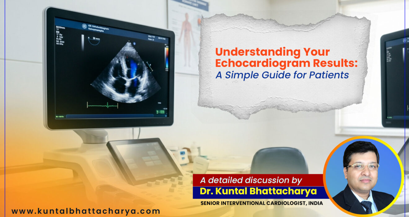
In simple terms, an echocardiogram (often referred to simply as an “echo”) is an ultrasound of your heart. Just as an obstetrician uses ultrasound to look at a baby inside the womb, a cardiologist uses the exact same soundwave technology to look at the structures of your beating heart.
A small, microphone-like device called a transducer is placed on your chest. It sends safe, painless, high-frequency sound waves into your body. When these sound waves hit your heart valves, muscles, and blood vessels, they bounce back (echo). The machine’s computer instantly translates these bouncing sound waves into moving, real-time pictures on a monitor.
Why Sound Waves Are Superior for Heart Imaging
Unlike X-rays or CT scans, an echocardiogram does not expose you to any harmful radiation. It is entirely non-invasive, meaning no needles or surgical cuts are involved. Furthermore, while an Electrocardiogram (ECG/EKG) measures the electrical activity of your heart, an echocardiogram shows the mechanical activity. It allows your doctor to see your heart actually squeezing and relaxing, to watch the valves open and shut, and to track the blood as it flows through the various chambers.
The Different Types of Echocardiograms
While the most common type is the Transthoracic Echocardiogram (TTE), which is done from the outside of the chest, there are other variations depending on what your doctor needs to see:
- Stress Echocardiogram: This involves taking ultrasound images before and immediately after you walk on a treadmill or ride a stationary bike. It helps doctors see how your heart muscle performs under physical stress.
- Transesophageal Echocardiogram (TEE): If the view from the outside of your chest is not clear enough (sometimes due to lung conditions or body tissue), a specialized tube with a tiny transducer is guided down your throat into your esophagus, which sits directly behind the heart. This provides highly detailed images of the heart valves.
- 3D Echocardiography: Advanced machines can now render a three-dimensional image of your heart, providing incredible depth and detail, which is particularly useful before valve surgeries.
Why Did Your Doctor Order This Test?
Investigating Unexplained Symptoms
If you have visited a clinic complaining of specific physical symptoms, an echocardiogram is usually the first major diagnostic tool deployed. We look for the root cause of:
- Shortness of Breath (Dyspnea): Especially if you find it hard to breathe when lying down flat or after mild physical exertion.
- Chest Pain (Angina): While chest pain can be caused by many things, an echo helps rule out structural heart problems.
- Palpitations or Irregular Heartbeats: Feeling like your heart is skipping a beat, fluttering, or racing.
- Swelling (Edema): Unexplained swelling in your legs, ankles, or abdomen can be a sign that your heart is not pumping blood efficiently, causing fluid to back up in your body.
- Heart Murmurs: If a doctor listens to your chest with a stethoscope and hears a “whooshing” or “swishing” sound, it usually indicates turbulent blood flow through a valve. An echo will identify exactly which valve is causing the noise.
Monitoring Existing Cardiac Conditions
If you have already been diagnosed with a heart issue, an echo is used as a routine tracking tool. For example, if you have had a heart attack in the past, we use the test to see how much of the heart muscle was damaged and if it is healing. If you are on medication for heart failure, the echo tells us if the medication is successfully improving your heart’s pumping strength.
Decoding the Medical Jargon: The Anatomy of Your Report
When you receive your printed report, it will likely be divided into several sections detailing the different structural parts of your heart. Let us break down the most critical metrics you will find on that piece of paper.
1. Left Ventricular Ejection Fraction (LVEF): The Pumping Power
This is often the most important number on your entire report. Your heart has four chambers: two upper (atria) and two lower (ventricles). The Left Ventricle is the main pumping chamber responsible for pushing oxygen-rich blood out to the rest of your entire body.
The Ejection Fraction (EF) is a percentage that represents how much blood the left ventricle pumps out with each and every contraction.
- Normal (50% to 70%): Do not panic if your number is not 100%. A normal, healthy heart does not empty entirely with each beat. An EF of 55% to 65% is considered perfectly healthy.
- Borderline (41% to 49%): The heart’s pumping ability is slightly below normal. This might be due to early-stage heart disease, a past minor heart attack, or uncontrolled high blood pressure.
- Reduced (Under 40%): This indicates that the heart muscle is weak and is not supplying enough blood to your body. This is a primary indicator of a condition known as heart failure. If your number is in this range, you must seek the guidance of a senior Interventional Cardiologist in Kolkata immediately to formulate a treatment plan.
2. The Heart Valves: The Doors of the Heart
Your heart has four valves (Aortic, Mitral, Tricuspid, and Pulmonary). Think of these valves as one-way doors that open to let blood pass through and snap shut tightly to prevent blood from flowing backward. Your report will note if there are any issues with these doors. The two main problems are:
- Stenosis (Stiffening): This means the valve flaps have become thick, stiff, or fused together. The door cannot open fully, which restricts the forward flow of blood. The heart has to work much harder to push blood through a narrowed opening.
- Regurgitation (Leaking): This means the valve does not close tightly. Like a leaky faucet, blood flows backward into the chamber it just left. You might see the words “Trace,” “Mild,” “Moderate,” or “Severe” regurgitation on your report. “Trace” or “Mild” is very common and often nothing to worry about. “Moderate” or “Severe” usually requires medical management or surgical repair.
3. Heart Chamber Sizes (Atria and Ventricles)
Your report will measure the physical dimensions of your heart chambers in millimeters or centimeters.
- Enlargement (Dilatation): If your report states that a chamber is “enlarged” or “dilated,” it means that part of the heart has stretched out and become larger than normal. This usually happens because the heart is trying to compensate for a leaky valve, high blood pressure, or a weak heart muscle. An enlarged heart has to work harder and is less efficient.
4. Heart Muscle Thickness (Hypertrophy)
Just like lifting weights makes your bicep muscles grow thicker, making your heart work too hard causes the heart muscle walls to thicken.
- Left Ventricular Hypertrophy (LVH): If your report notes LVH, it means the walls of your main pumping chamber are thicker than normal. The most common cause of this in India is chronic, untreated high blood pressure. While a thicker muscle sounds like a good thing, in the heart, it is not. Thick heart walls become stiff, making it difficult for the heart to relax and fill properly with blood between beats (this is often called “Diastolic Dysfunction” on your report).
5. Wall Motion Abnormalities
Your doctor looks at how evenly the heart muscle squeezes.
- Hypokinetic: Means a portion of the heart wall is moving weakly or sluggishly.
- Akinetic: Means a portion of the wall is not moving at all.
These terms usually indicate that a specific area of the heart is not getting enough blood flow due to a blocked coronary artery, or that the area was permanently damaged by a previous heart attack.
6. The Pericardium (The Heart’s Jacket)
The heart sits inside a thin, fluid-filled sac called the pericardium.
- Pericardial Effusion: If your report mentions this, it means there is excess fluid trapped in the sac surrounding the heart. A small amount of fluid can be normal or related to a viral infection, but a large amount can compress the heart and become a medical emergency.
The Procedure: What to Expect in Kolkata
Many patients feel anxious simply because they do not know what the hospital experience will be like. The standard Echocardiogram procedure kolkata is highly streamlined and comfortable.
How to Prepare
One of the best things about a standard Transthoracic Echocardiogram is that it requires absolutely zero preparation. You do not need to fast, and you can take all your regular medications with a normal glass of water. You should wear comfortable, two-piece clothing (like a shirt and trousers) because you will be asked to undress from the waist up and wear a hospital gown.
Inside the Examination Room
- You will be escorted to a dimly lit room (the low light helps the technician see the ultrasound screen more clearly).
- You will be asked to lie down on an examination table, usually on your left side. Lying on your left side brings your heart closer to the chest wall, allowing for clearer pictures.
- The sonographer (ultrasound technician) or cardiologist will apply a special water-based, hypoallergenic gel to your chest. This gel might feel a bit cold, but it is necessary to help the sound waves travel through your skin.
- The technician will firmly press the transducer wand against various spots on your chest, moving it around to capture the heart from multiple angles. You may feel slight pressure, but it should not be painful.
- You will hear loud “whooshing” or “pulsing” noises coming from the machine. Do not be alarmed! This is completely normal; the machine is simply translating the sound of your blood flowing into an audible noise (the Doppler effect).
- The technician may occasionally ask you to breathe in deeply, exhale, or hold your breath for a few seconds to get the best possible image.
- The entire process typically takes about 30 to 45 minutes from start to finish.
After the Procedure
Once the imaging is complete, the gel is wiped off with a towel, and you can get dressed immediately. You can drive yourself home and resume your normal daily activities, including eating and working, right away. There is no downtime or recovery period.
Let’s Talk Finances: Echocardiogram Cost in Kolkata
Healthcare expenses are a valid concern for many families. Fortunately, compared to more invasive cardiac tests, an echocardiogram is relatively affordable and highly accessible.
The Echocardiogram cost in Kolkata can vary depending on several factors, including the type of hospital (private vs. government), the locality, and the specific type of machine used (standard 2D vs. advanced 3D imaging).
- Standard 2D Echocardiogram: At top-tier private healthcare facilities like Narayana Health RN Tagore Hospital, the cost generally ranges from ₹1,500 to ₹3,500. Smaller diagnostic clinics may charge between ₹1,000 and ₹2,000.
- Stress Echocardiogram: Because this involves treadmill equipment and a longer observation time, the cost is slightly higher, usually ranging from ₹2,500 to ₹4,500.
- Transesophageal Echocardiogram (TEE): This is a specialized, semi-invasive procedure requiring mild sedation and specialized probes. The cost for a TEE generally starts from ₹4,000 and can go up to ₹8,000 or more.
It is always advisable to check with your health insurance provider, as routine diagnostic tests prescribed by a registered medical practitioner are often covered under standard health policies or corporate wellness benefits.
Taking Action: When Do You Need an Interventional Cardiologist in Kolkata?
Reading your report is only the first step; taking the appropriate medical action based on those results is what saves lives. If your echocardiogram reveals significant abnormalities—such as a low ejection fraction (under 40%), severe valve regurgitation, or akinetic wall motion—you must not ignore these red flags.
What to Do Next
- Do Not Self-Diagnose: The internet is filled with worst-case scenarios. Do not let Google induce panic. Medical reports must be interpreted in the context of your overall physical health, age, and medical history.
- Seek Specialist Consultation: General physicians are excellent for initial referrals, but complex heart issues require a specialist. You need an Interventional Cardiologist in Kolkata. Interventional cardiologists are specially trained to use minimally invasive, catheter-based techniques (like angioplasty, stenting, and pacemaker implantation) to treat cardiovascular disease without open-heart surgery.
- Gather Your Medical History: When you visit the specialist, bring your echocardiogram report, the CD containing the video images (if provided by the lab), your most recent ECGs, any recent blood test reports (especially Lipid Profile and HbA1c), and a complete list of your current medications.
Where to Go for Treatment
When dealing with the heart, the facility matters just as much as the doctor. You want to choose a hospital equipped with advanced Cath Labs, 24/7 cardiac emergency support, and a multidisciplinary team.
In Eastern India, Narayana Health RN Tagore Hospital (RTIICS) in Mukundapur, Kolkata, stands as a premier institution for comprehensive cardiac care. We are equipped with state-of-the-art infrastructure and a dedicated team capable of handling everything from routine checkups to the most complex structural heart disease interventions, including advanced coronary imaging, complex angioplasty, and TAVI/TAVR (valve replacement without open-heart surgery).
Finding the Right Adult Heart Doctor in Kolkata
Choosing the right physician is a deeply personal decision. Look for a doctor who:
- Has extensive, verifiable clinical experience and strong academic credentials (MBBS, MD, DM in Cardiology).
- Takes the time to explain your condition in simple terms rather than rushing you out of the office.
- Believes in a balanced approach—prioritizing evidence-based non-invasive medical management (lifestyle changes and medicines) first, and only recommending interventional procedures when absolutely necessary for your survival and quality of life.
Frequently Asked Questions (FAQs) About Echocardiograms
What is the difference between an ECG and an Echocardiogram?
Think of your heart like a house. An ECG (Electrocardiogram) checks the "electrical wiring" of the house—it looks for irregular heartbeats and arrhythmias. An Echocardiogram checks the "plumbing and structure" of the house—it looks at the walls, doors (valves), and the pumping power. Both tests complement each other, and doctors often order them together for a complete picture.
Can an echocardiogram detect if my arteries are blocked?
Not directly. An echocardiogram cannot see the inside of your coronary arteries to spot cholesterol blockages. However, it can detect the effects of a blockage. If a coronary artery is severely blocked, the heart muscle it supplies will not move normally (shown as hypokinetic or akinetic on the report). To actually see the blockages in the arteries, an Angiogram is required.
Are there any side effects or risks associated with the test?
For a standard Transthoracic Echocardiogram, there are absolutely no known risks, side effects, or radiation exposure. It is entirely safe for everyone, including pregnant women and children. If you undergo a Stress Echo, the risks are associated with the physical exercise on the treadmill (mild fatigue or a rare chance of triggering an irregular heartbeat), which is why a medical team monitors you the entire time.
Who is the best Echocardiogram doctor in Kolkata to evaluate my report?
A skilled sonographer performs the test, but the final interpretation must always be done by an experienced cardiologist. As a Senior Consultant at Narayana RN Tagore Hospital, I regularly evaluate complex echo reports to formulate personalized, life-saving treatment plans for my patients. You should seek out a specialist with years of hands-on clinical experience in both diagnostic and interventional cardiology.
How often should I get an echocardiogram done?
If your heart is completely healthy and you have no symptoms, you do not need annual echocardiograms. However, if you have a diagnosed heart condition (like heart failure, a valve defect, or a history of a heart attack), your cardiologist will likely recommend an echo once a year or every six months to monitor your progress and adjust your medications.
Why does my report say "Trace Regurgitation" in multiple valves? Should I be worried?
No, you should not be worried. It is incredibly common for healthy individuals to have "trace" or "physiologic" regurgitation (a minuscule leak) in the tricuspid or pulmonary valves. This is considered a normal variant of human anatomy and rarely progresses into a clinical problem. Your cardiologist will look at this and likely tell you your heart is functioning perfectly.
Conclusion: Your Heart is in Good Hands
Receiving a medical report filled with technical terminology can be a source of immense anxiety. However, knowledge is your strongest weapon in the fight against cardiovascular disease. Understanding the basic metrics of your echocardiogram—like your Ejection Fraction and the status of your valves—allows you to take an active, informed role in your own healthcare journey.
Remember, an abnormal result on an echocardiogram is not an immediate death sentence; it is a vital warning sign. It is the roadmap that tells us exactly what needs to be fixed. With the incredible advancements in modern medicine, many structural heart conditions and pumping deficiencies can be successfully managed with the right medications, lifestyle modifications, or minimally invasive procedures.
If you are currently holding a confusing report or experiencing unexplained chest pain or shortness of breath, do not wait for the problem to resolve itself. The heart is a resilient organ, but it requires timely care. Seek out an experienced Echocardiogram doctor in Kolkata for a proper clinical correlation.
Your heart has worked tirelessly for you since before you were even born. Take the necessary steps today to ensure it keeps beating strong for decades to come.
Disclaimer: The information provided in this blog post is for educational and informational purposes only. It does not constitute professional medical advice, diagnosis, or treatment. Always consult a qualified medical professional or cardiologist regarding any health concerns or before starting any new treatment plan.
Disclaimer: This blog post is for informational and educational purposes only and should not be considered a substitute for professional medical advice, diagnosis, or treatment. Always seek the advice of a qualified and experienced cardiologist in your area with any questions you may have regarding a medical condition.
Follow Dr. Kuntal Bhattacharya on Facebook & Youtube for regular updates.
Read Also:
- Life with an AICD: Managing Sudden Cardiac Death Risk Effectively
- How 3D Imaging Enhances Precision in TAVI Procedures in Kolkata
- Identifying Asymptomatic Heart Disease in Young Adults in 2026: The Silent Killer
- The Role of FFR in Deciding the Right Treatment for Heart Blockages
- Leadless Pacemakers vs. Traditional Pacemakers: Pros and Cons for 2026
- Managing Complex Coronary Artery Disease in 2026: A Cardiologist’s Guide
- How IVUS and OCT Technology Revolutionized Angioplasty Success in Kolkata
- Choosing the Right Coronary Angioplasty Doctor in Kolkata 2026: A Patient’s Guide
- Life After TAVI/TAVR Treatment: Patient Advice from a Cardiologist in Kolkata
- Why Choose TAVI/TAVR Over Open Heart Surgery in 2025?
- TAVI and TAVR Procedures in Kolkata in 2025
- Understanding TAVI / TAVR: The Future of Heart Valve Treatment
- Early Signs of Heart Valve Problems and When to Seek a TAVI Doctor
- Angioplasty vs. Bypass Surgery in 2025: What Should You Choose?
- Modern Technology in Cardiac Devices in 2025
- How to Find the Best TAVI and TAVR Doctor in Kolkata in 2025?
Stay Healthy. Stay Informed.